
What joint cartilage does
Articular cartilage provides a smooth, low-friction surface at the ends of bones. It has limited ability to repair itself after a substantial injury, particularly when the defect does not reach the underlying bone.
Cartilage layers and injury grades
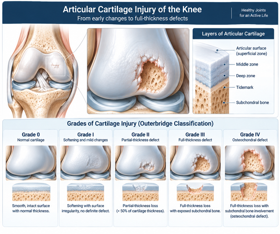
The infographic distinguishes the cartilage surface and deeper layers, then summarizes the Outerbridge grading spectrum from normal cartilage to a full-thickness osteochondral defect. Grading is one part of assessment and does not determine treatment by itself.
Focal cartilage injury
A focal defect is a localised area of damage surrounded by otherwise healthier joint surface. It can follow a twisting injury, impact or kneecap dislocation, and may cause activity pain, swelling, catching or a sense that the joint is not trustworthy.
Osteochondral lesions
An osteochondral lesion affects both cartilage and the bone beneath it. In some cases a fragment becomes unstable or loose in the joint. The stability, location, size and bone involvement affect urgency and treatment options.
Cartilage injury versus arthritis
A focal cartilage or osteochondral defect is a localised problem; arthritis is a broader, often progressive joint condition with diffuse cartilage loss, bone changes and sometimes deformity. Diffuse advanced arthritis is not treated by focal cartilage restoration. This distinction protects patients from an unrealistic procedure choice.
Symptoms and mechanical signs
Symptoms may include pain with impact or loaded bending, recurrent swelling, catching, stiffness or reduced confidence in sport. Symptoms alone cannot identify the lesion: similar complaints occur with meniscus tears, patellofemoral pain and arthritis.
Why alignment matters
Bow-legged or knock-kneed alignment can overload one compartment and threaten a repair. Long-leg standing X-rays may be used to assess the mechanical axis. In selected cases, correcting alignment is part of protecting the joint or a cartilage procedure.
Meniscus and ligament context
The meniscus distributes load, while stable ligaments guide movement. Meniscus deficiency or untreated instability can overload a cartilage defect. A complete plan considers whether these problems need rehabilitation, repair, reconstruction or other management alongside cartilage treatment.
Examination and plain X-rays
Examination assesses swelling, motion, joint-line tenderness, kneecap tracking, alignment and stability. Weight-bearing X-rays help show arthritis, alignment, bone changes and some osteochondral abnormalities; they may be normal in an early cartilage lesion.
The role of MRI
MRI helps define cartilage, subchondral bone, marrow oedema, loose bodies, menisci and ligaments. It is selected when it can answer a clinical question and is not necessary for every patient with knee symptoms. It is interpreted with symptoms and examination, because an imaging abnormality is not always the pain generator. CT may occasionally assist with detailed bone assessment or surgical planning.
Nonsurgical care
A period of diagnosis-specific nonsurgical care may include adjusting impact activity, physiotherapy for strength and movement control, maintaining fitness with lower-impact exercise, symptom-relieving medicines where appropriate and addressing body weight when relevant. This may improve symptoms even though it does not regrow normal cartilage.
Selected cartilage procedures
Selected patients with a symptomatic, contained focal lesion and an otherwise suitable joint may be considered for procedures such as marrow stimulation, osteochondral grafting or cell-based cartilage repair. The appropriate technique depends on lesion features, age, activity, bone involvement and concomitant alignment, meniscus or ligament issues.
Limits, recovery and expectations
Cartilage procedures involve a rehabilitation commitment and often restrictions on loading or impact for a period determined by the procedure. They cannot guarantee return to a particular sport or prevent future arthritis. Diffuse arthritis, major untreated malalignment or widespread joint damage may make restoration inappropriate.
When prompt review is needed
Seek timely assessment after a significant injury with inability to bear weight, a large swelling, deformity or a locked knee. A hot swollen knee with fever requires urgent assessment to exclude infection.
Can knee cartilage grow back on its own?
Substantial articular cartilage defects have limited spontaneous healing. Symptoms may improve with rehabilitation and load management, while selected focal defects may be considered for restorative procedures.
Is cartilage damage the same as arthritis?
No. A focal injury is localised; arthritis usually involves more widespread joint change. The distinction changes the likely benefit of cartilage restoration.
Will an MRI show all cartilage damage?
MRI is very useful but is interpreted alongside examination and X-rays. Its detail can vary with the lesion and scan quality.
Why are alignment X-rays needed?
They show how load passes through the leg. Malalignment may overload the damaged compartment and influence treatment planning.
Does every cartilage lesion need surgery?
No. Symptoms, lesion stability, joint health, activity goals and response to nonsurgical care determine whether surgery is considered.
Can cartilage restoration treat severe arthritis?
It is generally not designed to treat diffuse advanced arthritis. Other arthritis-focused approaches may be more appropriate after assessment.
Written and medically reviewed by: Dr. Yousef Abuodeh Consultant Orthopedic Surgeon Last medically reviewed: September 2, 2026