
Pelvic ring versus acetabulum
Pelvic-ring fractures affect the structural ring of the pelvis. Acetabular fractures involve the hip socket and its joint surface. Their implications, imaging needs and treatment planning differ.
High-energy versus fragility injuries
These fractures may follow major trauma, but they can also occur after a lower-energy fall in an older person or someone with osteoporosis. The mechanism and bone quality help shape assessment and treatment.
Associated injuries
Depending on the injury, there may also be abdominal or pelvic-organ, urinary-tract, neurological or vascular injuries, or fractures elsewhere. These do not occur in every case, but they are important considerations after significant trauma.
Initial assessment
Immediate trauma priorities, resuscitation and stabilization may take precedence over definitive orthopedic reconstruction. The initial plan is coordinated around the person’s overall condition and associated injuries.
X-rays and CT
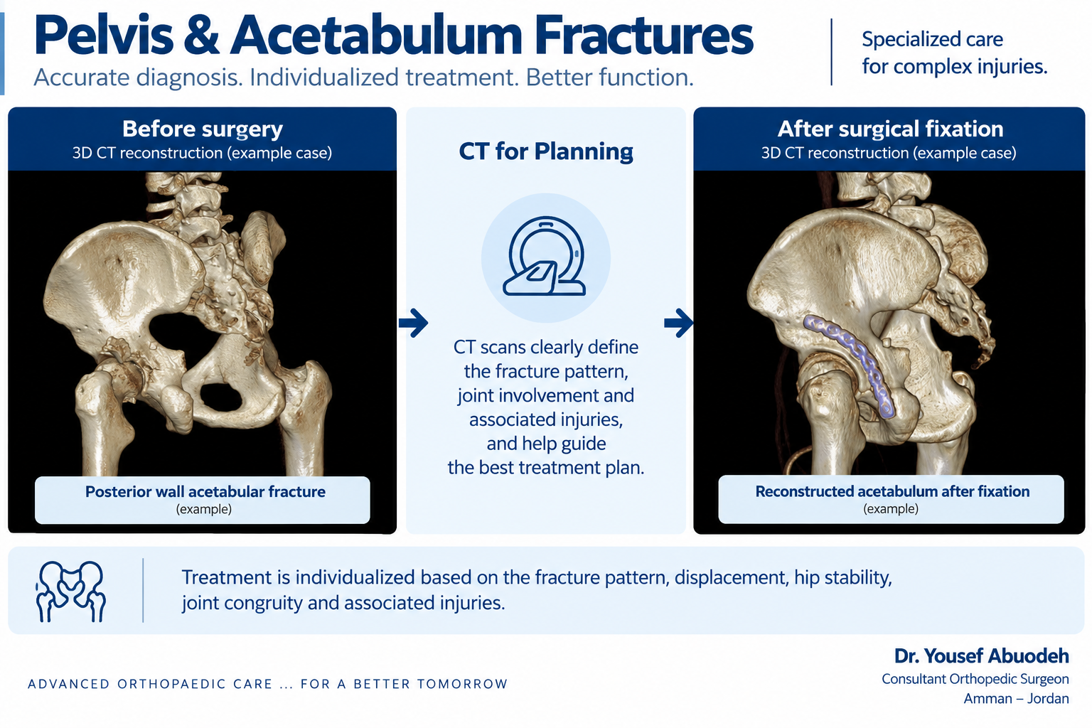
X-rays are part of the initial assessment in many cases. CT is important for many pelvic and acetabular fractures because it defines pattern, displacement and joint anatomy in greater detail, but imaging is selected according to the clinical situation.
Clinical example 1: posterior-wall acetabular fracture
This example demonstrates how three-dimensional CT can define a posterior-wall fracture pattern and assist individualized planning before surgical fixation.
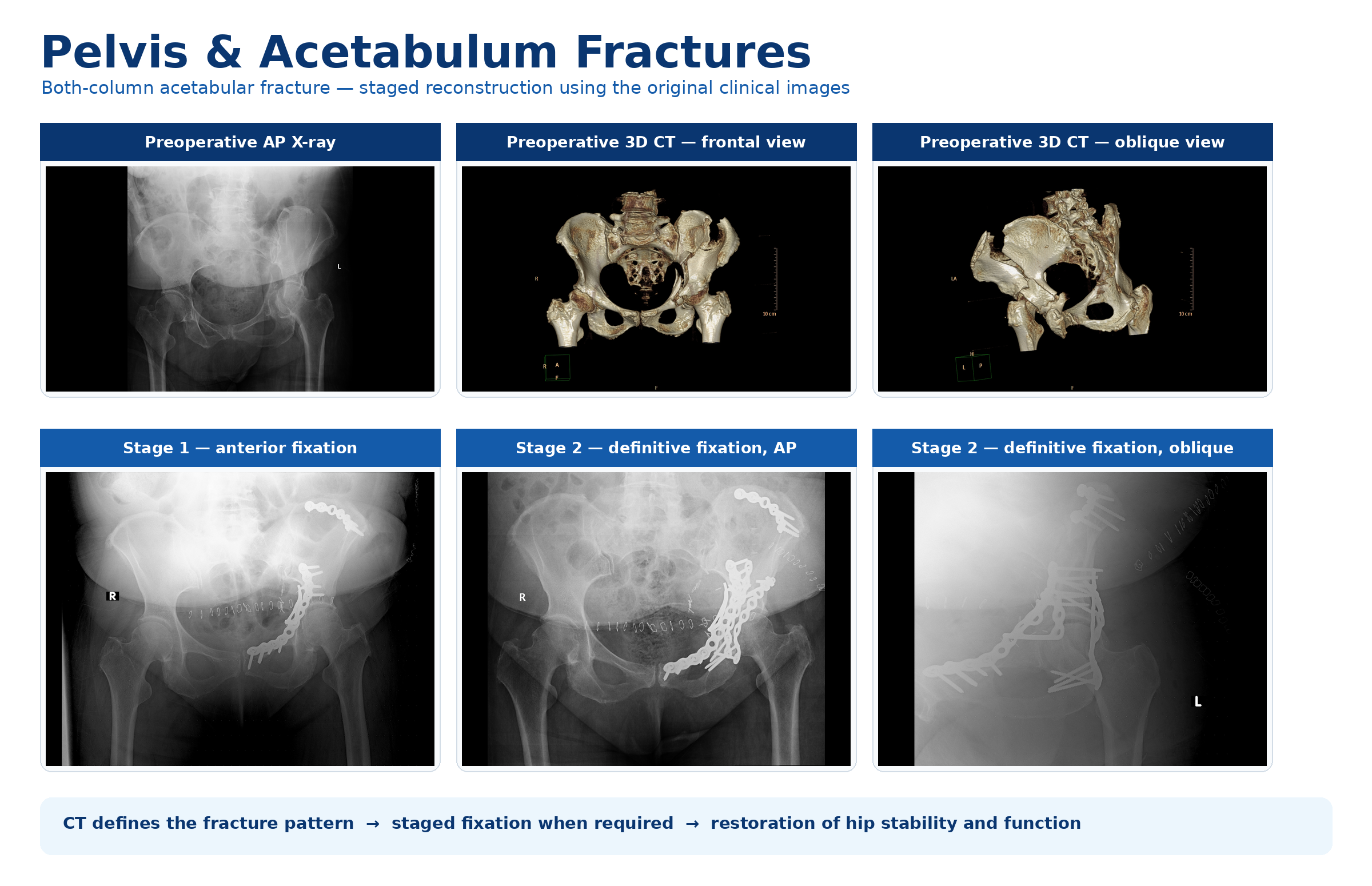
Clinical example 2: both-column fracture with staged reconstruction
This different clinical example shows preoperative radiographs and three-dimensional CT followed by staged reconstruction. It represents a separate fracture pattern and treatment sequence from the posterior-wall example above.
What determines nonsurgical versus surgical treatment?
The decision considers fracture pattern, displacement, stability, joint congruity, age, bone quality, overall health, functional demands and associated injuries. No single operation or approach suits every fracture.
Surgical planning
When surgery is appropriate, the strategy and approach depend on fracture anatomy, pattern, joint involvement and associated injuries. There is no universal surgical approach.
Weight bearing and rehabilitation
Restrictions vary and there is no universal timetable. Progression depends on fracture stability, healing and the treatment provided, with rehabilitation adjusted to the individual situation.
Urgent warning signs
Seek urgent care after significant trauma with severe pelvic or hip pain, inability to bear weight, major deformity, new neurological changes, urinary difficulty or blood in the urine, or symptoms such as faintness, marked weakness, pallor or rapid deterioration that may indicate instability or blood loss.
Why is CT important?
CT can define the fracture pattern, displacement and joint surface in more detail for planning, although imaging is selected according to the clinical situation.
Will I need surgery?
That depends on the individual injury, stability, displacement, joint congruity, associated injuries and health context after specialist assessment.
When can I bear weight?
Restrictions and progression are individualized according to fracture stability, healing and treatment; there is no single timetable for every injury.
Written and medically reviewed by: Dr. Yousef Abuodeh Consultant Orthopedic Surgeon